TYRION
Analysis of the photo
intraoperative
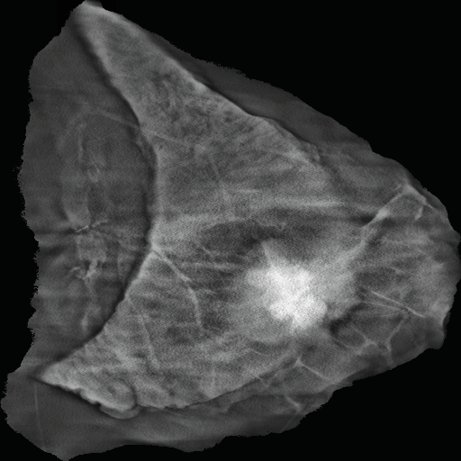
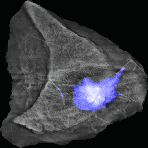
The basic functionality of the TYRION system is to perform an analysis of the intraoperative image, the result of which will be: the significance on the intraoperative X-ray of the tissue slice of the area occupied by the cancerous tumor, and the calculation of the excision margin – that is, the minimum distance of the tumor from the edge of the excision.
According to experts, the development of data and appropriate training of algorithms are the most important of the steps in the development of software to support medical imaging diagnostics. Working on real data, extracted from the clinical environment during actual surgeries, is absolutely crucial so that the finished system can be a support, not a distraction, for doctors.
To date, we have collected more than a hundred thousand images of breast tissue (individual X-ray scans included in the 3D tomosynthesis), which our team evaluated for quality and diagnostic value. Then, this data was marked by a team consisting of doctors and qualified specialists and rechecked by radiology experts to avoid any inconsistencies. In order to speed up the whole process and improve communication between the marker and the checking physician, we developed our own application based on machine learning algorithms, dedicated to the marking of x-ray images of breast tissue, distinguishing between tumor tissue and microcalcifications.